|
Services > Corneal
Disorders
|
|
|
Corneal
blindness accounts for 0.52% of total blindness in the
Indian subcontinent. The cornea can be the site of various
disorders. However before discussing the common problems,
which can affect the cornea, it is important to understand
the structure and function of the normal eye.
|
|
Normal
Structure & Function of the Eye
|
|
|
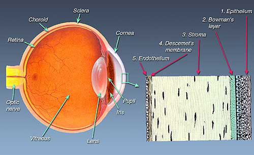
The
eyeball is made up of three "coats". The sclera ('white
of the eye') is the protective outer coat of the eye, the
front portion of which is transparent and is called the cornea.
The innermost lining or coat of the eyeball is the retina,
a thin light-sensitive film. Between the sclera and the
retina lies the choroid, which has a chiefly nutritive
function. Some distance behind the cornea is the iris,
which is visible as the 'colored portion of the eye' with
a central opening - the pupil. This is just like
the shutter or diaphragm aperture of the camera and helps
regulate the amount of light entering the eye. Behind the
iris lies the transparent lens, which helps in focusing
light on the retina. The space between the lens and the
retina is filled with a clear jelly called the vitreous
body. The
eye is like a camera in which lenses focus the picture on
a light sensitive film. In the human eye, the transparent
cornea and lens focus light on the retina, which changes
it into electrical signals, which are then transmitted to
the brain by the optic nerve to be perceived as images. |
|
How
is a cornea transplanted?
|
|
|
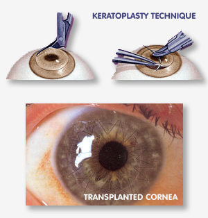
A
corneal transplantation, like a cataract operation, is
usually performed under local anesthesia. General anesthesia
is used for children and apprehensive or nervous patients.
The operation is completely painless and takes about one
hour to perform. The diseased, cloudy, opaque cornea is
removed from the recipient's (living patient's) eye, and
re placed by a new clear cornea (graft) from the donor's
(deceased person's) eye. The new cornea is then sutured
or stitched into place. As few as eight and as many as
20 or more sutures may be used, according to the size of
the graft, to hold the border of the graft to the border
of the recipient. If the operation is successful and the
graft "takes" and remains clear, the patient should see
well again, provided the lens and the retina behind the
cloudy cornea are normal.
The
patient is usually hospitalized for one day but requires
rest for the next one month although returning to light
work is not a problem. However frequent follow-ups are
required over the following six months to one year.
|
|
How
successful is corneal transplantation?
|
|
|
 In favorable subjects the rate of success of corneal
transplantation may be as high as 60%, with good final
visual acuity with glasses. In unfavorable subjects, the
rate of success may be around 10 to 20%. Each patient is
evaluated individually before definite results can be predicted. In favorable subjects the rate of success of corneal
transplantation may be as high as 60%, with good final
visual acuity with glasses. In unfavorable subjects, the
rate of success may be around 10 to 20%. Each patient is
evaluated individually before definite results can be predicted.
The
most important factors in determining the final results
are:
|
Basic
corneal disease (some types of corneal disease respond
better to corneal transplantation than others). |
|
State
of the donor's cornea. |
|
Surgical
technique and skill. |
|
Healing
ability of the recipient cornea. |
|
Sensitivity
reactions between donor and recipient cornea. |
A
corneal transplantation will not help every blind person
to see again. If a person is blinded by glaucoma, a detached
retina, or degenerative change and the retina has been
damaged or destroyed, nothing can restore lost sight.
Corneal transplantation restores vision only in eyes
that have been partially blinded by corneal disease.
Some vision must be present before transplantation is
even contemplated.
THE
OCCURRENCE OF PAIN, REDNESS, WATERING, LIGHT-SENSITIVITY
AND DIMINISHED VISION, ANY TIME (EVEN MONTHS OR YEARS)
AFTER CORNEAL TRANSPLANTATION SURGERY, REQUIRES IMMEDIATE
ATTENTION OF YOUR OPHTHALMOLOGIST. |
|
What
is Computer Vision Syndrome?
|
|
|
 Computer Vision Syndrome (CVS), a relatively new condition,
is the complex of eye and vision-related problems associated
with computer use affecting millions of people. The primary
symptoms are eyestrain, blurred vision, dry and irritated
eyes, tired eyes, and headaches. Neck and backaches can
also be related to the way that we use our eyes at the
computer. This happens because staring at a computer screen
causes a significant reduction of the normal blink rate.
Hence washing of the corneal surface of the accumulated
dust, debris and tear waste products is delayed; instead
they have a longer contact time with the cornea producing
ocular surfacing problems and eye fatigue. Computer Vision Syndrome (CVS), a relatively new condition,
is the complex of eye and vision-related problems associated
with computer use affecting millions of people. The primary
symptoms are eyestrain, blurred vision, dry and irritated
eyes, tired eyes, and headaches. Neck and backaches can
also be related to the way that we use our eyes at the
computer. This happens because staring at a computer screen
causes a significant reduction of the normal blink rate.
Hence washing of the corneal surface of the accumulated
dust, debris and tear waste products is delayed; instead
they have a longer contact time with the cornea producing
ocular surfacing problems and eye fatigue.
The
following steps can help alleviate your symptoms:
|
Lower
your computer screen so that the center of the screen
is 4-8 inches below your eyes. Correct posture, adequate
room lighting and convenient placement of the mouse
and keyboard, are essential to ensure comfort while
working on the computer. |
|
Use
an anti-reflection filter over your monitor to avoid
glare and eyestrain. |
|
If
you are seated in a draft or near an air vent, try
to eliminate the flow of air past your eyes. Low
humidity or fumes aggravate a dry eye condition.
If you have these conditions in your work place,
fix them if possible. |
|
Concentrate
on blinking whenever you begin to sense symptoms
of dry or irritated eyes. |
|
Every
once in a while (especially when you sense the symptoms)
close your eyes and roll them behind your closed
eyelids. Take a short break of a few minutes from
your work, every half an hour.
|
| |
Use
artificial tears to re-wet and lubricate your eyes.
Use as recommended by either your doctor or the manufacturer. |
You
should seek professional eye care if symptoms persist.
Many computer users need a pair of glasses for their computer
work that is different from the glasses they use for their
other common visual needs. They either have a different
prescription or a different lens design from their usual
glasses. A thorough check up by an ophthalmologist is essential
to identify and treat the factors contributing to the problem.
|
|
|